This factsheet is for people who are being tested for a heart condition, or who would like information about such tests.
A doctor may suggest one or more tests in order to understand the exact cause of symptoms such as chest pain or palpitations (an unpleasant awareness of your heartbeat, often described as thumping in your chest). Similar tests may be done for people who have had a heart attack.
About the heart
Your heart works as a pump, its muscular walls contracting to force the movement of blood. Each side of your heart is divided into an upper chamber (atrium) and a lower chamber (ventricle).
The heart, lungs and major vessels of the chest
Your heartbeat starts with an electrical impulse in your right atrium. This impulse causes the two atria to contract and pump blood into your ventricles through a set of heart valves. The electrical impulse carries on into your ventricles, causing them to contract, pumping blood around your body.
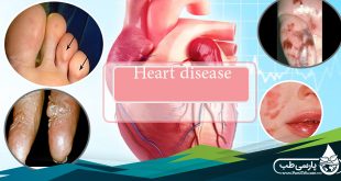
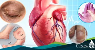
Your coronary arteries supply blood to your heart muscle. A common heart condition is coronary heart disease (CHD), in which fatty deposits (plaques) gradually block the coronary arteries. This can result in angina (a feeling of chest pain, chest tightness, and sometimes breathlessness or choking) or a heart attack.
Tests for heart conditions
Electrocardiogram (ECG)
An ECG is a test that measures the electrical activity of your heart to see how well it’s working.
Resting ECG
An ECG can often be done at your GP practice. It’s a simple test and only takes a few minutes. Small, sticky patches (electrodes) are put on your arms, legs, and chest. The electrodes are connected to a recording machine by wires. A trace (outline) of your heartbeat is shown on a screen or printed on to a paper strip.
The test can show if you have had or are having a heart attack, problems with heart rhythm, and enlargement or thickening of your heart.
This test won’t detect all heart disease and you may need to have other tests.
A man having a resting electrocardiogram
24-hour/seven-day ECG recording
This test continuously records an ECG over 24 hours or seven days.
Electrodes are put on your chest and connected by wires to a small electronic recorder that can be attached to your belt. You will need to note the time of any symptoms (such as palpitations) that you have during the test.
The test can help diagnose problems that only happen now and again, such as palpitations, or can show up an abnormal heart rhythm.
A man wearing a 24-hour electrocardiogram monitor
Cardiac event recorders
These devices record your heart’s activity over a longer time. They are used if you have symptoms that only happen occasionally, such as palpitations, dizziness, and fainting.
A portable event recorder is a device that you place over your heart when symptoms start. Pressing a button records an ECG.
An implantable loop recorder is a small, slim electronic box that is inserted beneath the skin of your chest (under local anesthesia) and left in place for up to 14 months.
Exercise ECG
This test records an ECG while you’re on a treadmill or exercise bike. The exercise starts off gently and is gradually made harder. It’s usually done in a hospital setting and lasts up to 15 minutes.
The test can show if your heart is getting enough blood through your coronary arteries when you’re active. This can help to show if you have CHD (although it isn’t always accurate). It can also assess the condition of your heart after a heart attack or after heart surgery.
A man on a treadmill having an exercise electrocardiogram
24-hour blood pressure recording
A small recorder worn on a belt around your waist is attached by wires to a cuff wrapped around your arm. A blood pressure reading is taken automatically about once every hour over a 24-hour period.
The test may be useful if your doctor thinks that your blood pressure is unusually high when measured in a medical setting.
Echocardiogram
This test uses ultrasound to produce a moving ‘real-time’ image of the inside of your heart. It’s done in an out-patient setting and can take up to one hour.
Harmless ultrasound waves are sent through your chest with a probe moved across your skin. The echoes that bounce off your heart are used to create a moving image of your heart on a screen.
The test provides information about the structure of your heart and how well it pumps. It may be used if you have recently had a heart attack or have heart failure (when your heart isn’t pumping as well as it should). It’s also used to assess the condition of your heart valves or if you have a problem with your heart present from birth (congenital heart disease).
Other forms of echocardiography include a transoesophageal echocardiogram (TOE) and fetal echocardiogram (done on an unborn baby).
In a TOE, the ultrasound probe is placed in your esophagus (the pipe that goes from your mouth to your stomach) to take the images of your heart. Your doctor will spray anesthetic onto the back of your throat before asking you to swallow a small ultrasound probe on the end of a thin, flexible tube. The probe can create images without your ribcage and lungs getting in the way.
A man having an echocardiogram
Radionuclide tests
These tests are also known as single-photon emission computed tomography (SPECT) or multiple-gated acquisition (MUGA) scans.
Myocardial perfusion scans
There are two parts to this test – rest and stress. It’s done in a hospital setting.
Your doctor will give you a small, harmless injection of radioactive material, which passes through your heart muscle. A large camera directed at your heart picks up rays sent out by the radioactive material and creates an image.
Your doctor will give you another injection and ask you to exercise on a treadmill or exercise bike. If you’re unable to do much exercise, your doctor may give you a medicine that makes your heart beat faster. The camera will create images of your heart, as before.
The test can help to diagnose CHD. It can check how well your heart is pumping and the flow of blood to your heart muscle.
Positron emission tomography (PET) scan
This test allows your doctor to see your heart working and examine the blood flow. It’s usually done in an out-patient setting and may take up to two and a half hours.
Your doctor will give you an injection of a small amount of radioactive material and will ask you to lie still under a scanning device.
The test is useful for deciding whether you need angioplasty (a procedure to widen a blood vessel by inflation of a balloon on the end of a catheter) or surgery if your condition is complicated.
Angiogram (cardiac catheterization)
This test looks inside your coronary arteries. It’s usually done as a day case, but you may need to recover overnight in the hospital. The test takes a minimum of half an hour and may take longer.
A long, thin, flexible tube (catheter) is put into a blood vessel in your groin and guided to your coronary arteries. A dye is injected through the catheter, which makes any narrowing or blockages in your coronary arteries clearly visible on an X-ray image. The test may be uncomfortable and there is some risk of complications.
The test shows if your coronary arteries have narrowed. It can also help your doctor decide on the type of treatment needed if you have CHD or angina. It can give information about the blood pressure inside your heart and how well it’s pumping.
Cardiac enzyme testing
During a heart attack, damaged heart muscle releases chemicals called enzymes into your blood. Levels of the enzymes can be measured by blood tests. The enzymes creatine kinase, troponin T, and troponin I are those most commonly tested for.
The test shows whether or not your heart muscle has been damaged.
Magnetic resonance imaging (MRI)
This test uses magnets and radio waves to produce images of the inside of your body. MRI doesn’t use X-rays. It’s done in an out-patient setting and takes about an hour.
Your doctor will ask you to lie still in a short tunnel, around which there is a large magnet, while the images are created.
The test shows the structure of your heart and the surrounding blood vessels. It can be useful for investigating CHD and cardiomyopathy (a condition when your heart muscle is diseased).
Electrophysiological (EP) testing
This test allows your heart’s electrical activity to be examined in detail. It may be done as a day case, but usually you need to recover overnight in the hospital. It usually takes two to three hours.
A catheter is put into a blood vessel in your groin and guided to your heart. The tip of the catheter stimulates your heart and records the electrical activity.
The test is used to diagnose abnormal heart rhythms and find out which part of your heart is affected.